
The epi-illumination fluorescence and bioluminescence imaging system was created for research studies with experimental animals and cell cultures in vivo using fluorescent and bioluminescent markers. The device allows noninvasive registering of processes in living organisms in real time. It can be used for solution of various scientific and technical problems related to the study of spatial distribution of fluorescent and bioluminescent substances in the objects under consideration.
A high sensitivity cooled EM CCD camera is used to detect low intensity bioluminescence. Superbright LED sources of visible range are used for fluorescence excitation. The radiation parameters correspond to the 1st class of laser hazards for skin and eyes of the operating staff.
General view of the device:
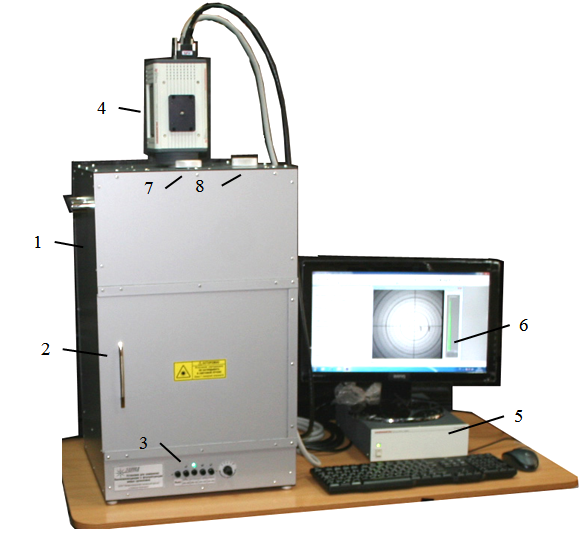 1 – dark chamber, 2 – sliding door of the dark chamber, 3 – control panel, 4 – cooled CCD camera, 5 – CCD camera power and control unit, 6 – PC monitor, 7 – cover of the window for installing optical filters, 8 – handle for selecting optical filter
Basic units and components layout:
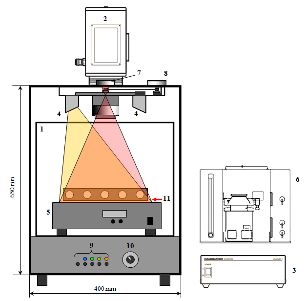 1 – dark chamber sliding door , 2 – cooled CCD camera, 3 – CCD camera power and control unit, 4 – superbright LED sources, 5 – platform of heating and anesthesia system for laboratory animals with gas stand with 5 lines of anesthetic agents delivery, 6 – gas tank of heating and anesthesia system, 7 – cover of the window for installing optical filters (“Filter change”), 8 – – handle for selecting optical filter (“Filter selection”), 9 – source selecting buttons, 10 – radiation intensity switch, 11 – plane of studied object location
Technical characteristics:
• CCD
– (optional) Hamamatsu EMCCD (monochrome, the number of pixels – 512х512, linear size of photosensitive area – 8.2х8.2 mm, quantum efficiency in the wavelength range of 500-700 nm is not less than 80%);
– possibility of installation of other CCDs on customer’s choice.
• Superbright LED sources
– nonuniform intensity distribution in the field of vision of the device not more than 15%;
– spectral characteristics on customer’s choice (not more than 5 various wavelength ranges).
• Emission optical filters
– spectral characteristics on customer’s choice (not more than 5 filters);
– possibility of installation of the automated filters change system (in development, expected by the end of 2014).
• Heating and anesthesia system
– (optional) used anesthetic — isoflurane, adjustable range of concentration —0%-5% in 0.5% increments, oxygen pressure at system entrance — 3.5 Atm. Preset temperature +37 0C, operating range of temperature setting from 33 0C to 40 0C with a step of 1 0C, temperature maintenance +2 0С.
Examples of obtained images:
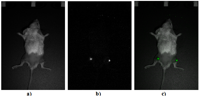 (a) “photo” of the surface of experimental mouse in scattered light, (b) bioluminescent sources in experimental mouse, (c) resultant composite image obtained by superimposing areas of the meaningful bioluminescence signal from image (b) on the photo of the object’s surface (a)