
Applications:
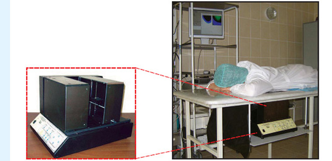
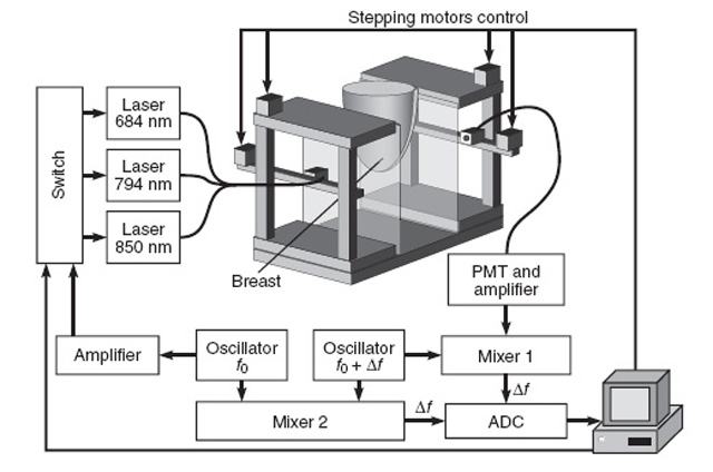
FD ODS setup was created to visualize neoplasia of breast tissue and to estimate its size. The breast is fixed between 2 glass plates and scans in transilluminative configuration using source-detector pair. At the source point we switch between 3 sources with different wavelengths (650, 794 and 850 nm). Spectral information is used for determination of tissue components, which is different in normal and cancer tissue.
Specifications:
– wavelengths 684, 794, 850 nm
– tissue thickness up to 80 mm
– amplitude modulation frequency, 140 MHz
FD ODS setup:
Scheme of FD ODS:
An example of the distribution of blood oxygen saturation level (%) in normal breast tissue (А) and in the case of breast cancer (В):
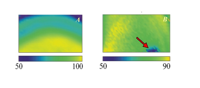 Image of normal breast tissue are characterized by homogeneity and absence of inclusions. In breast cancer, optical inhomogeneities are visualized; tumor zone is clearly defined against the normal tissues. Particularly, blood oxygen saturation level was lower in the projection of a tumor nodule. The arrow shows the tumor region.