
A fiber-based 525 nm laser generated 500 ps pulses with a 100–200 kHz repetition rate and an average power of 17 mW. An output laser fiber was coupled to the collimator with the rotating-beam homogenizer that aimed to provide the uniform illumination of the investigated object without speckles. The fluorescence excited in the investigated object was registered with the ICCD based on the MCP with the time resolution up to 200 ps.
Using ICCD the sequence of surface fluorescence images with different time delays relative to the exciting pulse was obtained. Based on these images, in each point of field, the lifetime estimated.
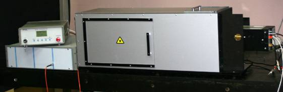
Setup:
Scheme:
The principle of data acquisition in the FLBI system using mise ex vivo with the 2.5 mm capsule containing DsRed2 FP implanted under the skin (Experiments were conducted in collaboration with the A.N. Bach Institute of Biochemistry RAS (Moscow, Russia)):
The results of in vivo whole-body lifetime imaging of subcutaneous TurboRFP-expressed tumors in nude mouse (Experiments were conducted in collaboration with the A.N. Bach Institute of Biochemistry RAS (Moscow, Russia)):
Please, contact us for detailed information
References:
1. Alexander L. Rusanov, Tatiana V. Ivashina, Leonid M. Vinokurov, Ilya I. Fiks, Anna G. Orlova, Ilya V. Turchin, Alexander P. Savitsky Life-time mode of FRET measurements for red fluorescent proteins. Journal of Biophotonics, 3(12), p. 774–783 (2010).