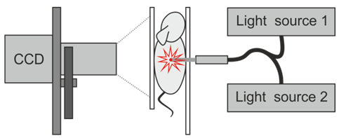
The device for transillumination fluorescence imaging is used for in vivo studies of 3D distribution of fluorescent markers inside experimental animals. The device operates in 3 regimes: epi-illumination, projection and diffuse fluorescence tomography (DFT).
Operating modes:
– The “epi-illumination“ (or back-reflection) mode enables fast (during a few seconds) assessment of the transversal size of fluorescent inclusion. It is realized with the use of superbright LED sources for fluorescence excitation, which provide uniform illumination of the experimental animal, and a CCD camera for registering object’s images through emission filters. The accuracy of assessment of the transverse dimensions of the fluorescent inclusion is the higher, the closer it is to the surface.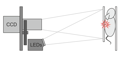
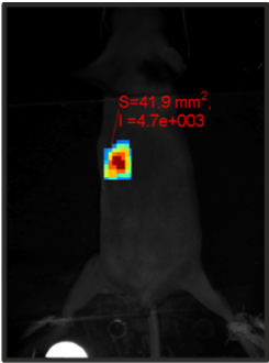 Image obtained in the epi-illumination mode. It shows the calculated area of the tumor S (in square millimeters) and its intensity I (in a.u.).
– The “projection“ mode (or transillumination raster scanning) is intended for studying experimental animals with deeply located fluorescent inclusions (such as, e.g., orthotopic fluorescent tumor). It is realized with the use of laser sources and a phomultiplier tube (PMT) as a detector with optical fibers and a mechanical 2D scanning system. The area of interest is selected on the experimental animal and scanned in 2D with selected scanning steps and integration time. Two-dimensional transillumination images of fluorescent inclusions are obtained as a result of scanning. It takes a few minutes (approx. 100 msec per point) to acquire the image.
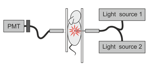
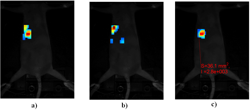 Combined images of fluorescent tumor obtained by scanning in the “projection” mode: a fluorescent transillumination image is obtained using light source 1 which excites fluorescence (a), normalization signal is obtained using light source 2 at the emission wavelength (b), normalized fluorescent image (c). The calculated tumor area S (in square millimeters) and its intensity I (in a.u.) are shown in the resulting composite image (c).
– The “DFT” mode (or 3D fluorescence imaging is now under development) is designed for studying objects with deeply located fluorescent inclusions. Allows reconstructing 3D images of the fluorophore distribution in the studied object by a set of different projections (measurements at multiple source and detector positions) of the object. This mode is similar to the “projection” mode with the only difference that a CCD camera with a filter bank is used as a receiver. This mode can be realized only at high signal-to-noise ratio, which is provided by using bright fluorophores whose spectra are within the transparency window (630-1000 nm). When fluorophores of the visible range (500-600 nm) are used, a highly sensitive CCD camera is required.
Technical characteristics:
• Light sources
– DPSS laser – 594nm wavelength for fluorescence excitation of far red dyes in the “projection” and “DFT” modes, optical fiber output (SMA 905 standard), amplitude modulation at a frequency of 2 kHz, average power on the surface of the studied object not more than 30 mW.
– Semiconductor laser for normalizing light propagating through the object in the “projection” and “DFT” modes – wavelength 642 nm, optical fiber output (SMA 905 standard), amplitude modulation at a frequency of 2 kHz, average power on the surface of the studied object not more than 15 mW.
– Superbright LED sources for fluorescence excitation in the “epi-illumination” mode – central wavelengths 467 nm, 518 nm, 590 nm, 635 nm (or on customer’s choice).
– Possibility of connection of one external laser source with intensity modulation by means of SMA connector.
• Emission optical filters
– Disk for installing filters – 6 positions, diameter of filters – 25mm.
– Spectral characteristics of filters – on customer’s choice.
• Mechanical scanning system
– Dual-axis scanning device – movement range 150x150mm, positioning accuracy on both axes not more than 0.2 mm.
• Photoelectron multiplier
– Type of photocathode – IR sensitized multialkaline
– Spectral range – from 300nm to 920nm
– Tuning range of PMT sensitivity – 1:104
• CCD
– monochrome, number of pixels – 1392х1040, linear size of photosensitive area – 10.2х8.3 mm, quantum efficiency in the wavelength range of 500-600 nm not less than 45%
• Dimensions
– Overall dimensions – 540х720х310mm (height x width x depth)
– Weight – 18 kg
General view of the device:
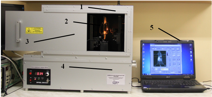 General view of the device with light protective case. 1 – dark chamber, 2 – experimental animal placed in a special transparent clamp, 3 – dark chamber door, 4 – apparatus unit, 5 – computer for controlling the device and data acquisition
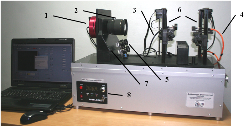 Device with removed light protective case. 1 – CCD camera, 2 – CCD camera lens, 3 – optical fiber bundle for radiation input into PMT unit, 4 – fiber output of laser sources 594nm, 642nm and of external laser source, 5 – superbright LED sources, 6 – electromechanical dual-axis scanning device, 7 – automated filter change unit, 8 – 594nm laser control unit.
